Showing posts with label Stem cell. Show all posts

Thursday, 30 January 2014

Groundbreaking Discovery : Embryonic Stem Cells Made With Acid

Stem cell 'major discovery' claimed


This is big.
Scientists have found a way to create embryonic stem cells without using an embryo or without introducing genetic material. The discovery could revolutionize medicine by giving doctors a way to repair diseased and damaged tissue — think heart disease, blindness, skin burns — with organs and tissue grown from the patient’s own cells.
Stem cell researchers are heralding a "major scientific discovery", with the potential to start a new age of personalised medicine.
Scientists in Japan showed stem cells can now be made quickly just by dipping blood cells into acid.
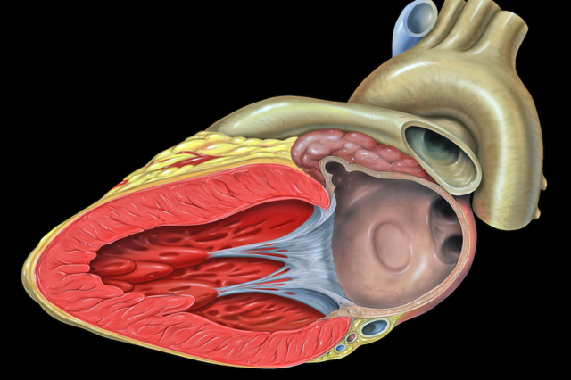
Stem cells can transform into any tissue and are already being trialled for healing the eye, heart and brain.
The latest development, published in the journal Nature, could make the technology cheaper, faster and safer.
The human body is built of cells with a specific role - nerve cells, liver cells, muscle cells - and that role is fixed.
However, stem cells can become any other type of cell, and they have become a major field of research in medicine for their potential to regenerate the body.
Embryos are one, ethically charged, source of stem cells. Nobel prize winning research also showed that skin cells could be "genetically reprogrammed" to become stem cells (termed induced pluripotent stem cells).

Acid bath
Now a study shows that shocking blood cells with acid could also trigger the transformation into stem cells - this time termed STAP (stimulus-triggered acquisition of pluripotency) cells.
Dr Haruko Obokata, from the Riken Centre for Developmental Biology in Japan, said she was "really surprised" that cells could respond to their environment in this way.
She added: "It's exciting to think about the new possibilities these findings offer us, not only in regenerative medicine, but cancer as well."
The breakthrough was achieved in mouse blood cells, but research is now taking place to achieve the same results with human blood.
Chris Mason, professor of regenerative medicine at University College London, said if it also works in humans then "the age of personalised medicine would have finally arrived."
He told the BBC: "I thought - 'my God that's a game changer!' It's a very exciting, but surprise, finding.
"It looks a bit too good to be true, but the number of experts who have reviewed and checked this, I'm sure that it is.
"If this works in people as well as it does in mice, it looks faster, cheaper and possibly safer than other cell reprogramming technologies - personalised reprogrammed cell therapies may now be viable."
For age-related macular degeneration, which causes sight loss, it takes 10 months to go from a patient's skin sample to a therapy that could be injected into their eye -and at huge cost.
Prof Mason said weeks could be knocked off that time which would save money, as would cheaper components.
'Revolutionary'
The finding has been described as "remarkable" by the Medical Research Council's Prof Robin Lovell-Badge and as "a major scientific discovery" by Dr Dusko Ilic, a reader in stem cell science at Kings College London.
Dr Ilic added: "The approach is indeed revolutionary.
"It will make a fundamental change in how scientists perceive the interplay of environment and genome."
But he added: "It does not bring stem cell-based therapy closer. We will need to use the same precautions for the cells generated in this way as for the cells isolated from embryos or reprogrammed with a standard method."
And Prof Lovell-Badge said: "It is going to be a while before the nature of these cells are understood, and whether they might prove to be useful for developing therapies, but the really intriguing thing to discover will be the mechanism underlying how a low pH shock triggers reprogramming - and why it does not happen when we eat lemon or vinegar or drink cola?"

2. New Method for Reprogramming Cells


Tuesday, 3 December 2013

For the first time, Human Stem Cells Converted to Functional Lung Cells

Human Stem Cells Converted to Functional Lung Cells


For the first time, scientists have succeeded in transforming human stem cells into functional lung and airway cells. The advance, reported by Columbia University Medical Center (CUMC) researchers, has significant potential for modeling lung disease, screening drugs, studying human lung development, and, ultimately, generating lung tissue for transplantation. The study was published today in the journal Nature Biotechnology.

"Researchers have had relative success in turning human stem cells into heart cells, pancreatic beta cells, intestinal cells, liver cells, and nerve cells, raising all sorts of possibilities for regenerative medicine," said study leader Hans-Willem Snoeck, MD, PhD, professor of medicine (in microbiology & immunology) and affiliated with the Columbia Center for Translational Immunology and the Columbia Stem Cell Initiative. "Now, we are finally able to make lung and airway cells. This is important because lung transplants have a particularly poor prognosis. Although any clinical application is still many years away, we can begin thinking about making autologous lung transplants -- that is, transplants that use a patient's own skin cells to generate functional lung tissue."
The research builds on Dr. Snoeck's 2011 discovery of a set of chemical factors that can turn human embryonic stem (ES) cells or human induced pluripotent stem (iPS) cells into anterior foregut endoderm -- precursors of lung and airway cells. (Human iPS cells closely resemble human ES cells but are generated from skin cells, by coaxing them into taking a developmental step backwards. Human iPS cells can then be stimulated to differentiate into specialized cells -- offering researchers an alternative to human ES cells.)
In the current study, Dr. Snoeck and his colleagues found new factors that can complete the transformation of human ES or iPS cells into functional lung epithelial cells (cells that cover the lung surface). The resultant cells were found to express markers of at least six types of lung and airway epithelial cells, particularly markers of type 2 alveolar epithelial cells. Type 2 cells are important because they produce surfactant, a substance critical to maintain the lung alveoli, where gas exchange takes place; they also participate in repair of the lung after injury and damage.
The findings have implications for the study of a number of lung diseases, including idiopathic pulmonary fibrosis (IPF), in which type 2 alveolar epithelial cells are thought to play a central role. "No one knows what causes the disease, and there's no way to treat it," says Dr. Snoeck. "Using this technology, researchers will finally be able to create laboratory models of IPF, study the disease at the molecular level, and screen drugs for possible treatments or cures."
"In the longer term, we hope to use this technology to make an autologous lung graft," Dr. Snoeck said. "This would entail taking a lung from a donor; removing all the lung cells, leaving only the lung scaffold; and seeding the scaffold with new lung cells derived from the patient. In this way, rejection problems could be avoided." Dr. Snoeck is investigating this approach in collaboration with researchers in the Columbia University Department of Biomedical Engineering.
"I am excited about this collaboration with Hans Snoeck, integrating stem cell science with bioengineering in the search for new treatments for lung disease," said Gordana Vunjak-Novakovic, PhD, co-author of the paper and Mikati Foundation Professor of Biomedical Engineering at Columbia's Engineering School and professor of medical sciences at Columbia University College of Physicians and Surgeons.

Via Science daily


Monday, 2 December 2013

First use of retrograde gene therapy on a human heart

First use of retrograde gene therapy on a human heart

Procedure delivers stem cells to the heart to repair damaged muscle and arteries
December 2, 2013
JuventasRetrograde_11-04-13a
Dr. Patel performing the operation on patient Ernie Lively on Nov. 7 (credit: University of Utah)
A new procedure designed to deliver stem cells to the heart to repair damaged muscle and arteries in the most minimally invasive way possible has been performed for the first time by Amit Patel, M.D., director of Clinical Regenerative Medicine and Tissue Engineering and an associate professor in the Division of Cardiothoracic Surgery at the University of Utah School of Medicine.
Patel started investigating cell and gene-based therapies for the treatment of heart disease 12 years ago, but only recently received FDA approval to try the therapy on Lively, who was the first of several patients anxious to receive the treatment.
More than 6 million people are currently living with heart failure. As the condition progresses, patients’ options are usually limited to a heart transplant or assist devices, such as an artificial heart. Patel wanted to find a way to intervene in the progression of heart failure before a patient advanced to the point of needing a heart transplant or device.
A minimally invasive surgical procedure
Patel and his team came up with the idea of retrograde heart therapy, a concept that has been discussed for 50 years. The first successful procedure was performed Nov. 7 on actor Ernie Lively, whose credentials that include a long list of TV and film appearances, including Passenger 57 and The Sisterhood of the Traveling Pants.
“It’s incredible. Imagine having a heart procedure that can potentially regenerate or rejuvenate your heart muscle — and it’s done as an outpatient procedure,” said Patel.
Patel uses a minimally invasive technique where he goes backwards through a patient’s main cardiac vein, or coronary sinus, and inserts a catheter. He then inflates a balloon in order to block blood flow out of the heart so that a very high dose of gene therapy can be infused directly into the heart.
The unique gene therapy doesn’t involve viruses (a rarity for gene therapy, Patel notes) and is pure human DNA infused into patients.  The DNA, called SDF-1, is a naturally occurring substance in the body that becomes a homing signal for a patient’s body to use its own stem cells to go to the site of an injury.
Once the gene therapy is injected, the genes act as “homing beacons.”  When the genes are put into patients with heart failure, they marinate the entire heart and act like a look out, he said.
When the signal, or the light from the SDF-1, which is that gene, shows up, the stem cells from not inside your own heart and from those that circulate from your blood and bone marrow all get attracted to the heart which is injured, and they bring reinforcements to make it stronger and pump more efficiently,” said Patel.
Patel said watching Lively recover successfully from the surgery is both rewarding and exciting for what the future holds for the procedure and those who may benefit from it.   He is already training other physicians around the U.S. to model what he accomplished first this month. He is overseeing a trial of the procedure in which 72 patients will participate over the next few months.
“This is one of the great moments in biological therapy for the heart,” said Patel. “We are providing options for patients who have no possible solutions. This is one of the safest and most reproducible therapies out there for these very sick patients.”
“He saved my life,” said Lively.

Wednesday, 27 November 2013

Scientists grow artificial skin from stem cells of umbilical cord


Researchers made a new type of artificial skin using stem cells from the umbilical cord, fibrin and agarose. This is great news because this type of skin can be stored in tissue banks and can be applied instantly, speeding the recovery process of burn victims. 

Read more: http://bit.ly/IrlDEc via Medical News Today

Friday, 25 October 2013

Scientist have discovered how the brain balances the number of stem cell & neuron during development


One of the major processes of brain development is the differentiation of neural stem cells into neurons and glia. It's a tricky process, because the brain also needs to make sure the stem cells don't proliferate out of control. In a new study published in Cell Reports, researchers at the University of Southern California identified a protein called SMEK1 that promotes stem and progenitor cell differentiation. But the scientists found that SMEK1 also works with a second protein known as Protein Phosphatase 4 to suppress neurogenesis. Only when new neurons are no longer being born can neural stem cells differentiate into neurons and glial cells like astrocytes and oligodendrocytes. Neural stem cell treatment has been proposed for several neurodegenerative diseases, and understanding stem cell differentiation could help scientists better harness their power.

Read more: http://bit.ly/1a5AWcc
Journal article: Protein Phosphatase 4 and Smek Complex Negatively Regulate Par3 and Promote Neuronal Differentiation of Neural Stem/Progenitor Cells. Cell Reports, 2013.

Wednesday, 4 September 2013

Mending a Broken Heart? Scientists Transform Non-Beating Human Cells Into Heart-Muscle Cells

Photo

For the first time lab grown human heart tissue beats on its own 

 In the aftermath of a heart attack, cells within the region most affected shut down. They stop beating. And they become entombed in scar tissue. But now, scientists at the Gladstone Institutes have demonstrated that this damage need not be permanent -- by finding a way to transform the class of cells that form human scar tissue into those that closely resemble beating heart cells.

Last year, these scientists transformed scar-forming heart cells, part of a class of cells known as fibroblasts, into beating heart-muscle cells in live mice. And in the latest issue of Stem Cell Reports, researchers in the laboratory of Gladstone Cardiovascular and Stem Cell Research Director Deepak Srivastava, MD, reveal that they have done the same to human cells in a petri dish.

"Fibroblasts make up about 50% of all cells in the heart and therefore represent a vast pool of cells that could one day be harnessed and reprogrammed to create new muscle," said Dr. Srivastava, who is also a professor at the University of California, San Francisco, with which Gladstone is affiliated. "Our findings here serve as a proof of concept that human fibroblasts can be reprogrammed successfully into beating heart cells."
In 2012, Dr. Srivastava and his team reported in the journalNature that fibroblasts could be reprogrammed into beating heart cells by injecting just three genes, together known as GMT, into the hearts of live mice that had been damaged by a heart attack. They reasoned that the same three genes could have the same effect on human cells. But initial experiments on human fibroblasts from three sources -- fetal heart cells, embryonic stem cells and neonatal skin cells -- revealed that the GMT combination alone was not sufficient.
"When we injected GMT into each of the three types of human fibroblasts, nothing happened -- they never transformed -- so we went back to the drawing board to look for additional genes that would help initiate the transformation," said Gladstone Staff Scientist Ji-dong Fu, PhD, the study's lead author. "We narrowed our search to just 16 potential genes, which we then screened alongside GMT, in the hopes that we could find the right combination."

In regenerative medicine breakthrough, lab-grown human heart tissue beats on its own